The order Teloschistales (with nearly 2,000 known species and accounting for > 10% of all known lichen-forming fungi) is one of the three main orders recognized within the largest subclass of the Lecanoromycetes in the Lecanoromycetidae. This subclass represents the core of the lichen-forming fungal diversity.
Interestingly, Xanthoria parietina (Teloschistales, Teloschistaceae) was the first lichen-forming species selected for genome sequencing. Lichen sexual reproduction is directly associated with fungal meiosis leading, for most lichens, to the formation of ascospores, and requiring the finding of the appropriate algal or cyanobacterial partner; i.e., horizontal transmission of the photobiont from generation to generation of lichens. For example, all the orange or dark brown to black disks shown in Fig. 1 are ascomata producing ascospores. Sexual reproduction is the dominant reproduction mode of lichens, however, many lichens can disperse their mycobiont and photobiont simultaneously within specialized propagules (e.g., soredia; Fig. 2) or through thallus fragmentation; i.e., vertical transmission of the photobiont. Lichens reproducing with these bi-partite asexual propagules usually don’t reproduce sexually.
Currently, the order Teloschistales encompasses two suborders, the Teloschistineae (composed of the three families Letrouitiaceae, Megalosporaceae, and Teloschistaceae) and the Physciineae, which contains the Caliciaceae (a predominantly buellioid clade of 731 species; Fig. 1G) and Physciaceae (a rinodinoid clade of 510 species; Fig. H-I). The recognition of this group of lichens at the ordinal level, and the inclusion of these five families within this order, are now broadly accepted.
The delimitation of the Caliciaceae and Physciaceae remains unclear to this day. Shared phenotypic similarities by members of these two families and several molecular studies support a close relationship. The Letrouitiaceae (Fig. 1E) is a monotypic family (Letrouitia), with about 15 species. Members of this family are widely distributed in subtropical and tropical regions, and are mostly corticolous. The Brigantiaeaceae, with 25 species and two genera, Argopsis and Brigantiaea, are found on bark in the tropics and on soil or decaying vegetation in the Arctic.
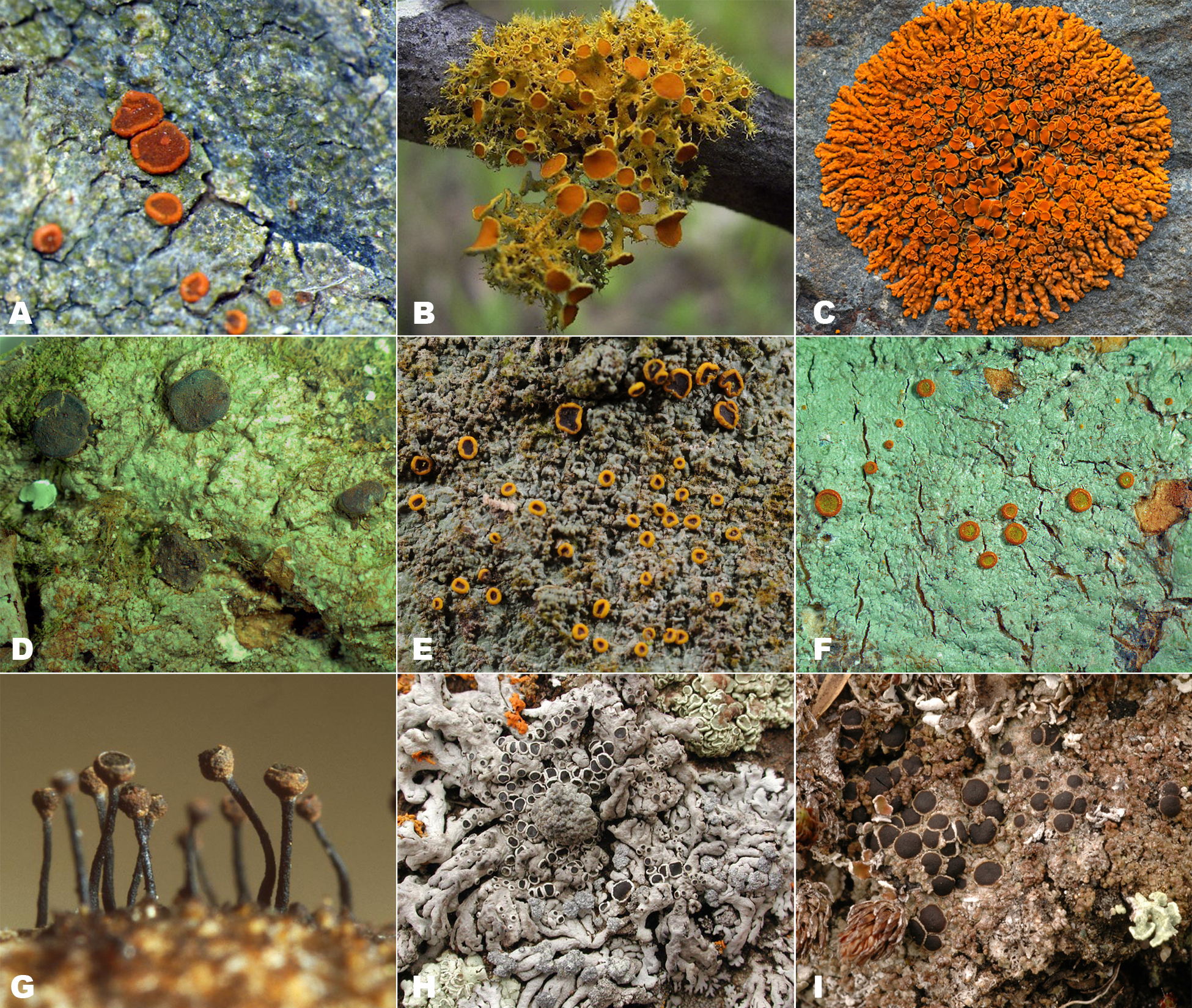 |
Figure 1. A. Caloplaca ceracea (Teloschistaceae); crustose endolithic growth-form. B. Teloschistes chrysophthalmus (Teloschistaceae); fruticose. C. Xanthoria elegans (Teloschistaceae); foliose. D. Megalospora tuberculosa (Megalosporaceae); crustose. E. Letrouitia domingensis (Letrouitiaceae); crustose. F. Brigantiaea leucoxantha (Brigantiaeaceae); crustose. G. Calicium salicinum (Caliciaceae); ascomata close-up, crustose thallus. H. Physcia phaea (Physciaceae); foliose. I. Rinodina mniaraea (Physciaceae); crustose. Photos by P. Whelan (A), D. Mosquin (C), E. Gaya (D and F), E. Timdal (E, H, I), and R. Haugan (G).
The Megalosporaceae (c. 39 species; Fig. 1D) includes three genera (Megalospora, Megaloblastenia and Austroblastenia) that are also mostly found in the tropics. The family Teloschistaceae (Fig. 1A-C) is composed of 11 genera and approximately 650 species (Caloplaca [c. 510 spp.], Cephalophysis [1 sp.], Fulgensia [8 spp.], Ioplaca [2 spp.], Josefpoeltia [3 spp.], Seirophora [11 spp.], Teloschistes [33 spp.], Xanthodactylon [1 sp.], Xanthomendoza [18 spp.], Xanthopeltis [1 sp.] and Xanthoria [56 spp.]). The traditional taxonomy of the family was based on vegetative features of the thallus, such as growth form (Fig. 1A-C) and presence/absence of a lower cortex, and occasionally on secondary substance composition. Initially, polarilocular ascospores were thought to be a diagnostic trait for this family, but with the inclusion of other genera such as Cephalophysis, Fulgensia and Xanthopeltis, which have simple or septate spores, the main features defining the family had to be reconsidered. The delimitation among genera included within the Teloschistaceae is highly artificial and in need of revision. No phylogenetic study to date has been designed to sample a balanced representation of taxa across the Teloschistales, i.e., Physciineae and Teloschistineae, to confront the current taxonomic delimitations at the family and genus levels. Moreover, we have reached the limit of the resolving power that a single-locus or two-locus phylogenetic studies selected from the nuclear ribosomal tandem repeat can provide within this order. In this context, an extensive taxon sampling within the Teloschistales with more loci (especially nuclear protein-coding genes) are needed for major advancements to be made in their classification and our understanding of macroevolutionary trends within this order. Additionally, this project is a case study of the new array of primers developed by AFTOL 2 for the systematic community.
The Teloschistaceae are collectively a model system to reconstruct the evolution of lichen growth forms because all main growth forms (including a major reduction of the thallus [Fig. 1A], and most intermediate forms) are found, and seem to be evolving quickly, in this family (Fig. 1A-C). Lichens are also known to produce a wide variety of unique secondary compounds, especially polyketide derivatives, terpenes and pulvinic acid derivatives. These chemical compounds are of ecological, taxonomic and evolutionary importance. Hence, another specific goal of our first main objective focusing on the families within the Teloschistales is to better understand the evolution of their secondary compounds. Members of the Teloschistales (and the Teloschistaceae alone) are found growing on all main types of substrates lichens are known to colonize, such as rocks, soils, mosses, dead wood and bark of living woody plants. This exhaustive study of the Teloschistales was designed to reconstruct and date the transitions from one substrate to another and determine how these transitions relate to the evolution of land plants. Our nearly complete lack of understanding of how these three major traits evolved, linked to the absence of a comprehensive phylogenetic framework and systematic phenotypic characterization of each major clades has been impeding our ability to change the classification the Teloschistales, which has been known to be flawed for more than half a century.
The reproductive biology of lichens has rarely been studied. The genetic mechanism responsible for lichen-forming ascomycetes to either reproduce sexually (Fig. 1) or to disperse by means of specialized vegetative symbiotic propagules containing both symbionts (Fig. 2) is unknown. Yet, these different types of reproduction determine if the photobiont is transmitted horizontally (sexual reproduction of the mycobiont) or vertically (asexual reproduction with vegetative symbiotic propagules) from generation to generation. Lichen-forming fungal species are usually mostly committed to reproduce sexually (e.g., Fig. 1) or asexually (Fig. 2), suggesting a genetic switch being involved. One key factor governing the sexual reproductive mode in fungi is the presence/absence of so-called mating-type (MAT) genes. In obligate outcrossing (heterothallic) species two nonallelic versions of the locus, termed idiomorphs, are found in isolates with complementary mating types. In contrast, facultative selfing (homothallic) species usually contain a single MAT locus with both mating types. The evidence cumulated so far support the expectation that asexual reproduction through specialized symbiotic propagules is restricted to obligate outcrossing heterothallic individuals of lichens. The impact of changes in mating systems (i.e., from heterothallism to homothallism) on speciation has been studied in some Teloschistaceae. Preliminary studies on lichens support the current view that homothallism is always the derived state, as the most common and widespread Teloschistaceae turned out to be homothallic. This unidirectional model of evolution has never been tested within a phylogenetic framework.
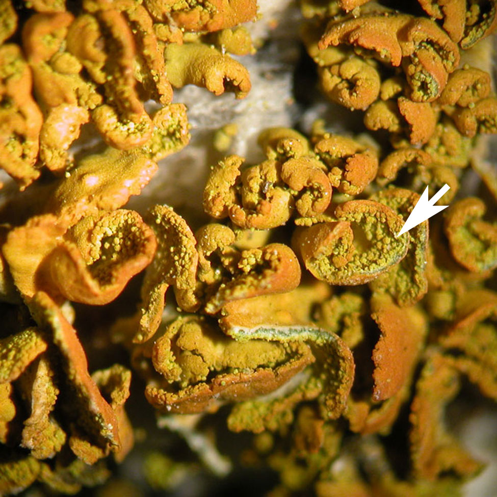 |
Figure 2. Lichen (Xanthomendoza mendozae) reproducing asexually with abundant specialized propagules (soredia [arrow]) containing both symbionts. Photo by C. Wagner.